

The Challenges of Interpreting Neck X-Rays in Horses

In the next instalment of my Sue's Snippets of Science series of articles I take a closer look at the challenges of radiological interpretation of the caudal cervical and cranial thoracic regions and discuss what is ‘normal’?

An abnormality on an X-ray does not necessarily mean that a horse is in pain or has a current or future problem. For example, X-ray changes that look like osteoarthritis do not always cause pain. Improvements in radiographic equipment now allow general equine vets to take cervical (neck) X-rays, but interpreting them can still be difficult. The images must be correctly exposed, the horse must stay still, and both the neck and X-ray beam must be properly aligned. If the image is angled or oblique, accurate interpretation may not be possible. Achieving all of this is often easier said than done.
Because X-rays of the cervical and cranial thoracic regions are now easier to take in the field, suspected neck problems are being diagnosed more often. However, many mature, comfortable horses have X-ray changes in the cervical vertebrae that cause no clinical signs, especially in the lower part of the neck.
Vets and other professionals may sometimes link signs such as poor hindlimb power to X-ray changes in the lower neck without proof that the two are connected. Other conditions can also look like neck problems. For example, a horse may resist turning to the right and feel stiff through the neck, when the real problem is right hindlimb lameness. Once the lameness is removed with appropriate nerve blocks, the horse may turn easily even though X-rays still show changes in several neck vertebrae. In other words, an X-ray abnormality doesn't automatically explain the horse’s clinical signs.
It's therefore essential to recognise the clinical features that may point to a primary neck problem and to decide which X-ray findings are likely to matter. It's equally important to recognise changes that are unlikely to be clinically significant. Bones change in size and shape in response to how they are loaded and stressed (Wolff’s law). The lower part of a horse’s neck has a wide range of movement: compare a horse lifting its head over a stable door, raising it before or after a jump, and lowering it to graze. It's therefore unsurprising that modelling changes, meaning changes in the size and shape of the articular processes, are common. These changes do not necessarily mean that osteoarthritis is present.
Learn more with Sue
Congenital Variants of Cervical Vertebrae
For more than 100 years it's been recognised that there are congenital (present at birth) anatomical variations in the shape of the caudal cervical vertebrae.
The variations are:
The absence of one or both of the ventral laminae of the transverse processes of the sixth cervical vertebra.
The presence of one or both ventral laminae on the ventral aspect of the seventh cervical vertebra (Compare Figures 1 and 2 below).

3. Occasionally, the first rib may be hypoplastic (small) or absent on one or both sides.
4. There may also be asymmetry in size of the left and right caudal cervical and cranial thoracic parts of the longus colli muscle which extends from the ventral aspect of the first cervical to the sixth thoracic vertebrae.
It has been suggested that this muscle asymmetry may make the vertebrae less stable and increase the risk of cervical abnormalities, including osteoarthritis of the articular process joints, with a wide range of possible clinical signs.1
There has been considerable debate about whether these congenital variants are clinically important and whether they are linked to other X-ray changes. Earlier studies found the variants of the sixth and seventh cervical vertebrae in about 30%–35% of different horse populations, but their relationship with clinical signs was unclear.2-7 It seems unlikely that an X-ray feature found in such a large proportion of horses would be clinically important in every case; that would not make biological sense. Even so, the term equine complex vertebral malformation (ECVM) has become increasingly common in the equestrian world.
Is The Presence of Congenital Variants of Likely Clinical Significance?
So, is ECVM actually a problem? The term ‘equine complex cervical malformation’ was introduced by non-veterinarians. The condition was compared with complex vertebral malformation syndrome in Holstein calves, a lethal inherited autosomal recessive disease in which calves die young and often have congenital heart abnormalities. However, the two conditions are not alike: affected calves have not been reported to have changes in the ventral laminae of the cervical vertebrae.
It has been also proposed by non-veterinarians that the acquisition of radiographs of the caudal aspect of the neck should be a routine part of pre-purchase examinations and that horses with these congenital variants should not be recommended for purchase.
A Large Scale Two Centre Case Control Study
A recent large collaborative study by UC Davis in California and the Animal Health Trust in the UK examined whether these congenital variations were linked to clinical conditions. The prospective study included Warmblood horses aged 3 to 22 years.8 Every horse had X-rays of the lower neck, from the fifth cervical to the second thoracic vertebra, regardless of its clinical history. The images were assessed using a detailed, pre-agreed protocol, and the assessors did not know whether each horse was a case or a control. All horses received the same thorough clinical assessment. Most were examined on the lunge and ridden when it was safe to do so, with further tests guided by the signs observed. The study included 96 cases: horses with incoordination and weakness consistent with spinal cord compression in the caudal cervical–cranial thoracic region, neck-related forelimb lameness, or neck pain and/or stiffness. It also included 127 control horses. These were either undergoing a pre-purchase examination or had a problem shown to be unrelated to the neck, such as hindlimb lameness that disappeared after diagnostic anaesthesia.
The ventral profile of the seventh cervical vertebra was abnormal in 24% of horses (Fig. 2). These horses had congenital transposition of one or both ventral laminae, a proportion similar to earlier studies.9 Importantly, cases were significantly less likely than control horses to have congenital variants. There was also no association between a congenital variant of the seventh cervical vertebra and modelling—changes in shape and size—of the articular processes from the sixth cervical to the second thoracic vertebra.
Congenital variants of the seventh cervical vertebra were not linked to any other X-ray findings, including spondylolisthesis, where adjacent vertebrae are partly displaced or misaligned (Fig. 3). In this group of horses, the congenital variants were therefore unlikely to be clinically important.

There was no association between the presence or absence of a congenital variant and the type of case. This reinforces the observation that these congenital variants are unlikely to contribute in any way to neck pain or stiffness, neck-related forelimb lameness or incoordination and weakness in most horses.
Some horses with congenital variants of the sixth and seventh cervical vertebrae may also have uneven development of the longus colli muscle. However, many other large muscles surround the neck vertebrae evenly on all sides. These muscles are likely to provide enough stability despite the congenital variants.
The congenital variants involving the ventral laminae of the transverse processes of the sixth and seventh cervical vertebrae are not comparable with the lethal inherited complex malformation found in Holstein calves. For this reason, the term ‘equine complex vertebral malformation’ (ECVM) should be avoided, because it may be misleading.
Cases were more likely than control horses to have severe modelling, changes in shape and size graded as none, mild, moderate or severe—of the articular processes between the sixth and seventh cervical vertebrae, or between the seventh cervical and first thoracic vertebrae.9,10
Mild and moderate modelling were common in both cases and controls, so these findings must be interpreted carefully (Fig. 4). Severe changes in the size and shape of the articular processes may restrict movement, cause pain, compress a nerve root and contribute to forelimb lameness, or compress the spinal cord and cause incoordination (Fig. 5). Some, but not all, horses with severe modelling also had osteoarthritis.


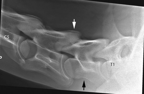
Cases were more likely than control horses to have spondylolisthesis, meaning partial displacement or misalignment, between the sixth and seventh cervical vertebrae or between the seventh cervical and first thoracic vertebrae (Fig. 6). Changes involving the intervertebral discs, with or without changes in the neighbouring vertebral bodies — known as discospondylosis—were also more common in cases than in control horses. These changes affected joints from the sixth cervical to the second thoracic vertebrae (Fig. 7).


The results show that horses can have a wide range of X-ray abnormalities without any related clinical signs. This underlines the need for a careful clinical assessment: an abnormal X-ray does not, by itself, prove that a horse has a neck-related problem.
Horses with neck-related signs may have X-ray abnormalities involving the first and second thoracic vertebrae, either on their own or alongside changes elsewhere (Figs. 6 and 7). If imaging stops at the cervical vertebrae and does not include the first two thoracic vertebrae and the cranial ribs, important lesions may be missed. A complete X-ray assessment of the cervicothoracic region is therefore important for an accurate diagnosis and prognosis, and for planning treatment and management.
Overall, congenital variants of the sixth and seventh cervical vertebrae were less common in cases than in control horses. They were not linked to other X-ray abnormalities that can restrict movement, cause pain, or affect the spinal cord or nerve roots. In the horse populations studied, congenital variants of the ventral laminae of the transverse processes of the sixth and seventh cervical vertebrae were therefore unlikely to be clinically important.
Horses in Germany Undergoing a Pre Purchase Examination
Another recent study found no link between congenital variants and competition performance. It included 200 Warmblood dressage and show jumping horses that had pre-purchase examinations between 2020 and 2024.11 Competition results came from the German Equestrian Federation database. One or both ventral laminae of the transverse processes of the sixth cervical vertebra were absent in 30% of horses. Modelling of the articular processes between the sixth and seventh cervical vertebrae was graded as absent, mild, or moderate–severe. Mild modelling was present in 26.5% of horses, while only 6% had moderate–severe modelling.
Horses with a congenital variant (23.3%) were less likely to have modelling of the articular processes between the sixth and seventh cervical vertebrae than horses with a normal sixth cervical vertebra (36.4%). There was no association between congenital variants and competition results. This adds to the evidence that, in most horses, congenital variants of the sixth and seventh cervical vertebrae are unlikely to be clinically significant.
Conclusions
Many horses have X-ray abnormalities in the lower cervical and upper thoracic vertebrae. These findings must be interpreted carefully and considered alongside the horse’s clinical signs. In most horses, a congenital variant of the ventral laminae of the sixth cervical vertebra is unlikely to be clinically important. However, abnormalities of the first ribs may occasionally be linked to clinical signs.
Acknowledgements:
Professor Monica Aleman, principal collaborator.
An anonymous donor to the Equine and Comparative Neurology Research Group, UC Davis, California, USA.
Learn more with Sue:
References
1. May-Davis S, Walker C. Variations and implications of the gross morphology in the longus colli muscle in thoroughbred and thoroughbred derivative horses presenting with a congenital malformation of the sixth and seventh cervical vertebrae. J Equine Vet Sci. 2015;35:560–568.
2. De Rouen A, Spriet M, Aleman M. Prevalence of anatomical variation of the sixth cervical vertebra and association with vertebral canal stenosis and articular process osteoarthritis in the horse. Vet Radiol Ultrasound. 2016;57:253–258.
3. Veraa S, De Graaf K, Wijnberg I, Back W, Verbgooij H, Neilen M, Belt A. Caudal cervical vertebral morphological variation is not associated with clinical signs in Warmblood horses. Equine Vet J. 2020;52:219-224. doi: 10.1111/evj.13140
4. Santinelli I, Beccati F, Arelli R, Pepe M. Anatomical variations of the spinous and transverse processes in the caudal cervical and the first thoracic vertebrae in horses. Equine Vet J. 2016;48:45-4
5. Veraa S, Bergmann W, Van den Belt A-J, Wijnberg I, Back, W. Ex vivo computed tomographic evaluation of morphology variations in equine cervical vertebrae. Vet Radiol Ultrasound. 2016;57:482–488. doi: 10.1111/vru.12393
6. Beccati F, Pepe M, Santinelli I, Gialletti R, Di Meo A, Romero J. Radiographic findings and anatomical variations of the caudal cervical area in horses with neck pain and ataxia: case control study on 116 horses. Vet Rec. 2020; doi:10.1136/vr.105756
7. Gerlach K, Schad P, Offhaus J, Brehm W, Pelli A. Incidence of variations of the transverse processes of the sixth and seventh cervical equine vertebrae. Pferdeheilkunde. 2021;37:605–610. doi:10.21836/PEM20210606
8. Dyson, S., Zheng, S., Aleman, M. Primary phenotypic features associated to caudal neck pathology in Warmblood horses. J. Vet. Int. Med. 2024, 38, 2380-2390. doi: 10.1111/jvim.17125
9. Dyson, S., Phillips, K., Zheng, S., Aleman, M. Congenital variants of the ventral laminae of the sixth and seventh cervical vertebrae are not associated with clinical signs or other radiological abnormalities of the cervicothoracic region in Warmblood horses. Equine Vet J 2025, 57: 419-430. doi: 10.1111/evj.14127
10. Dyson, S., Quiney, L., Phillips, K., Zheng, S., Aleman, M. Radiological abnormalities of the cervicothoracic vertebrae in Warmblood horses with primary neck-related clinical signs compared with control horses. Vet Radiol Ultrasound 2024, 65: 755-768. doi: 10.1111/vru.13420
11. Strootmann, T., Peter, V., Körner, J. Radiographic prevalence of anatomical variations of the ventral lamina of the sixth cervical vertebra, C6/C7 articular process joints modelling and competition outcomes in Warmblood sport horses. Animals 2026, 16: 424.


An excellent summary of an area which is a minefield and very often misinterpreted using the relevant literature.