
Exploring Equine Anatomy: A Visual Journey at the Horses Inside Out Exhibition
- Jessica Limpkin

- Apr 11, 2024
- 6 min read
Updated: Apr 25, 2025
This year as part of the Horses Inside Out conference at Loughborough University in February, there was the opportunity to visit Gillian Higgins’ Anatomy Exhibition, either as part of the whole weekend experience, or as a stand alone entry for the exhibition alone, and it was bigger and more comprehensive than ever.
Attracting visitors from around the world, this unique educational experience delved into the world of equine anatomy with a particular focus on growth and development. All the hours of planning and hard work, and the building of equine skeletons was worth it. The exhibition showcased the intricate structures that lie beneath the skin and how they differ in horses from young to old.


“Fascinating and educational. As a visual learner, it makes such a difference to see the exhibits first hand. Life-size and with the time to view and analyse them. What an incredible, unique learning opportunity.” Clare Chamberlayne BHSI
I had heard a few rumours about some of the additions to this year’s exhibition and was so excited to see it. This year's exhibition had an 2 additional whole horse skeletons, one of which was fully put together in a canter pose. So, in total, there were 6 full skeletons of different ages, sizes and all displayed in different poses.
This was all on top of the previous exhibition pieces some of you may have seen at the conference before, which include the full skeleton of Gillian’s own horse, the original Horses Inside Out horse ‘Freddie Fox’, who was unveiled at the 2023 conference (see video below), models of the Sacroiliac region, the hock and the carpus and many other fascinating models both hand made and real bone specimens. And the display of Scientific Posters, this year all related to studies in Growth & Development (the theme of this years conference). As well as that, there were talk zones with other professionals. Chris Pearce, Equine Vet & Dental Specialist led the "Head Zone" while Mark Johnson, Farrier & Hoof Care Professional was in charge of the "Leg Zone".
The exhibition was split into sections - the skeleton zone, the leg zone, the head zone and the art zone and in this article I will share with you what I learnt and appreciated most in each - so read on!!
Following the success and popularity of this exhibition Gillian and her team will be hosting another Equine Anatomical Art Exhibition this summer open from 22 – 25 August 2024, 10am-4pm at Horses Inside Out, Wavendon Grange, Old Dalby, LE14 3LW. I am already looking forward to it!
The Skeleton Zone

The most recent skeleton addition was a 3 yr & 4 month old Welsh Sec D mare, that had sadly been euthanised following chronic laminitis and had been donated to Gillian for the exhibition. I was fascinated to see this skeleton, firstly as I have a Welsh Sec D myself, and getting the see the size & scale of the bones in a horse similar to my own was incredible.
But also to have the opportunity to study the bones and the growth plates, and see just how developed (or not) a horse is at this age, an age at which many of us are starting to ride & train our horses.
"I’ve had shivers down my spine, literally as I look at the skeleton of a young horse taken by laminitis and now, serving as yet another phenomenal educational specimen being displayed this year. Rising 4 this horse and so many like it lack skeletal maturity, you can see so many un-fused growth plates and an amazing immature spinous process with for heaven sake being opaque enough for light to shine through. Surely we have to keep banging this message out to give time to horses to properly mature. Just because it looks big, strong and healthy, it doesn’t necessarily equate to what’s beneath the skin. I defy anyone lucky enough to be attending this anatomy exhibition not to learn something or think differently in some way." Mark Johnson

"Growth and development truly is a journey of a lifetime. Riders often think of horses as either immature or mature. LouLou gives a great insight into "partial maturity" and helps us to appreciate which areas are still skeletally immature in young horses when we first start riding them." says Gillian

Another addition to the exhibition was a foal skeleton, supplied by hoof care practitioner Siobhan Dillon. Again, fascinating to see and compare to the 3yr old mare.
With the 12yr old pony Skeleton and Freddie having been in his 20’s it was possible to see the whole range of bone development as well as bony changes that develop as well as other pathologies.

There was also the opportunity in the exhibition to label the bones on one of the skeletons and to put a pony skeleton together, as well as build a hock and a carpus joint. You could have spent hours in there being interactive with the bones and the other visitors having discussions.
The Leg Zone

Looking at the feet of the Welsh D was also really interesting, if a little troubling, a stark example of what laminitis can do.
It highlighted to me again just how important management is in the prevention of problems. If conditions are less than ideal and symptoms not attended to quickly enough the effects of laminitis can be catastrophic.
Mark Johnson, Farrier & Hoof care Professional was in charge of the "Leg Zone" and as well as Gillian's lower limb freeze-dried specimens, models of the sacroiliac ligaments and iliopsoas muscle group, Mark’s table had a vast array of preserved legs & feet, showing bone alignment in the leg, pedal bone positioning and what the same foot can look like with and without a balanced trim.
Mark was able to offer valuable insight into the anatomy of the foot and I was so pleased to be able to study and discuss these anatomical specimens.

The Head Zone

Gillian is well-known for her anatomical paintings to help make learning easier. In the head zone was an incredible painted model created by Gillian as well as 3D anatomical head models which were great for gaining a better understanding of the external anatomy structures of the head.
They gave a detailed insight into the delicate structures of the horse’s head. Highlighting the muscles, nerves and blood vessels which we must all strive to have a clearer understanding of to help us when it comes to bridle fit, reading facial expression, manual therapy and making sure the horse is as comfortable as possible.

There were also a set of models provided by Somso illustrating the horses’ teeth at various ages. During Dr Chris Pearce's talk he discussed how to age a horse by looking at the teeth so to have these models as well as the skulls and teeth in the skeleton zone gave a real opportunity to practice this skill and I really enjoyed studying these.
Equine Anatomical Art by Gillian Higgins

The equine anatomical art section included exciting new 3D models painted by Gillian herself depicting the muscles and skeleton in Gillian's usual style as well as some with a clever twist on word-art with the skeleton and muscles illustrated using the names of the bones written in the shape and location to depict the skeleton. What a fun way to learn the names of the structures.


I am in awe of some of the art created by Gillian, which is not only beautiful but also insightful and can be used as a learning tool. Some very lucky people got to take home the word art ponies, and statement canvases as well as anatomical posters. What a great fun way to display equine anatomy and spark conversation & intrigue
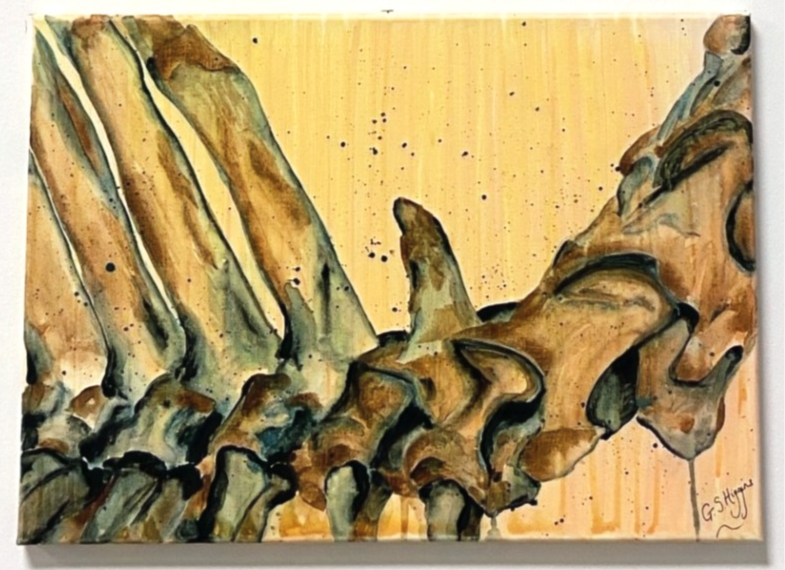

Gillian has depicted the anatomy of the horse from a number of different perspectives to get us all thinking about the horse in different ways and there really is something to spark the imagination of everyone whatever your equestrian background.
Gillian has drawn inspiration from different artistic styles from impressionist, abstract to pop art as well as a number of famous anatomists and equestrian artists including: George Stubbs, Leonardo da Vinci, and one of her latest collections inspired by Eadweard Muybridge is the "Skeletons in Motion" Print Collection.
If you like the look of these check out the prints and posters in the Horses Inside Out Shop:
Jess


Comments